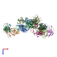
3hei

X-ray diffraction
2Å resolution

Ligand Recognition by A-Class Eph Receptors: Crystal Structures of the EphA2 Ligand-Binding Domain and the EphA2/ephrin-A1 Complex

Released:

Function and Biology Details

Structure analysis Details

Assembly composition:
hetero dimer (preferred)
PDBe Complex ID:
PDB-CPX-149073 (preferred)
Entry contents:
2 distinct polypeptide molecules
Macromolecules (2 distinct):
Ephrin type-A receptor 2 Chains: A, C, E, G, I, K, M, O
Molecule details ›
Chains: A, C, E, G, I, K, M, O
Length: 174 amino acids
Theoretical weight: 19.91 KDa
Source organism: Homo sapiens
Expression system: Homo sapiens
UniProt:
  • Canonical: P29317 (Residues: 28-201; Coverage: 18%)
Gene names: ECK, EPHA2
Sequence domains: Ephrin receptor ligand binding domain
Structure domains: Galactose-binding domain-like
Ephrin-A1, secreted form Chains: B, D, F, H, J, L, N, P
Molecule details ›
Chains: B, D, F, H, J, L, N, P
Length: 132 amino acids
Theoretical weight: 15.88 KDa
Source organism: Homo sapiens
Expression system: Homo sapiens
UniProt:
  • Canonical: P20827 (Residues: 18-147; Coverage: 69%)
Gene names: EFNA1, EPLG1, LERK1, TNFAIP4
Sequence domains: Ephrin
Structure domains: Cupredoxins - blue copper proteins

Ligands and Environments

No bound ligands
No modified residues

Experiments and Validation Details

Entry percentile scores
X-ray source: APS BEAMLINE 24-ID-C
Spacegroup: P1
Unit cell:
a: 58.483Å b: 102.108Å c: 135.255Å
α: 84.25° β: 79.77° γ: 73.94°
R-values:
R R work R free
0.175 0.172 0.233
Expression system: Homo sapiens