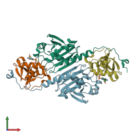
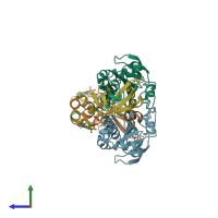
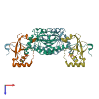
3hy2

X-ray diffraction
2.1Å resolution

Crystal Structure of Sulfiredoxin in Complex with Peroxiredoxin I and ATP:Mg2+

Released:

Function and Biology Details

Reactions catalysed:
Thioredoxin + ROOH = thioredoxin disulfide + H(2)O + ROH
Peroxiredoxin-(S-hydroxy-S-oxocysteine) + ATP + 2 R-SH = peroxiredoxin-(S-hydroxycysteine) + ADP + phosphate + R-S-S-R
Biochemical function:
Cellular component:

Structure analysis Details

Assembly composition:
hetero tetramer (preferred)
PDBe Complex ID:
PDB-CPX-170402 (preferred)
Entry contents:
2 distinct polypeptide molecules
Macromolecules (2 distinct):
Peroxiredoxin-1 Chains: A, B
Molecule details ›
Chains: A, B
Length: 206 amino acids
Theoretical weight: 23.06 KDa
Source organism: Homo sapiens
Expression system: Escherichia coli
UniProt:
  • Canonical: Q06830 (Residues: 1-199; Coverage: 100%)
Gene names: PAGA, PAGB, PRDX1, TDPX2
Sequence domains:
Structure domains: Glutaredoxin
Sulfiredoxin-1 Chains: X, Y
Molecule details ›
Chains: X, Y
Length: 110 amino acids
Theoretical weight: 11.99 KDa
Source organism: Homo sapiens
Expression system: Escherichia coli
UniProt:
  • Canonical: Q9BYN0 (Residues: 32-137; Coverage: 77%)
Gene names: C20orf139, SRX, SRX1, SRXN1
Structure domains: Conserved hypothetical protein from pyrococcus furiosus pfu- 392566-001, ParB domain

Ligands and Environments

2 bound ligands:
No modified residues

Experiments and Validation Details

Entry percentile scores
X-ray source: RIGAKU MICROMAX-007
Spacegroup: P212121
Unit cell:
a: 57.33Å b: 92.41Å c: 131.85Å
α: 90° β: 90° γ: 90°
R-values:
R R work R free
0.226 0.224 0.273
Expression system: Escherichia coli