3j2t

Electron Microscopy
9.5Å resolution

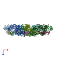
An improved model of the human apoptosome

Released:
Source organisms:
Primary publication:
Changes in Apaf-1 conformation that drive apoptosome assembly.
Biochemistry 52 2319-27 (2013)
PMID: 23521171
Related structures: EMD-5186

Function and Biology Details

Structure analysis Details

Assembly composition:
hetero tetradecamer (preferred)
Assembly name:
PDBe Complex ID:
PDB-CPX-127068 (preferred)
Entry contents:
2 distinct polypeptide molecules
Macromolecules (2 distinct):
Apoptotic protease-activating factor 1 Chains: A, B, C, D, E, F, G
Molecule details ›
Chains: A, B, C, D, E, F, G
Length: 1263 amino acids
Theoretical weight: 144.1 KDa
Source organism: Homo sapiens
Expression system: Spodoptera frugiperda
UniProt:
  • Canonical: O14727 (Residues: 1-1248; Coverage: 100%)
Gene names: APAF1, KIAA0413
Sequence domains:
Cytochrome c Chains: H, I, J, K, L, M, N
Molecule details ›
Chains: H, I, J, K, L, M, N
Length: 104 amino acids
Theoretical weight: 11.6 KDa
Source organism: Bos taurus
UniProt:
  • Canonical: P62894 (Residues: 2-105; Coverage: 99%)
Gene names: CYC, CYCS
Sequence domains: Cytochrome c

Ligands and Environments

2 bound ligands:
No modified residues

Experiments and Validation Details

Entry percentile scores
Resolution: 9.5Å
Relevant EMDB volumes: EMD-5186
Expression system: Spodoptera frugiperda