


Function and Biology Details
Reactions catalysed:
H(2)CO(3) = CO(2) + H(2)O
Urea = cyanamide + H(2)O
Biochemical function:
Biological process:
Cellular component:
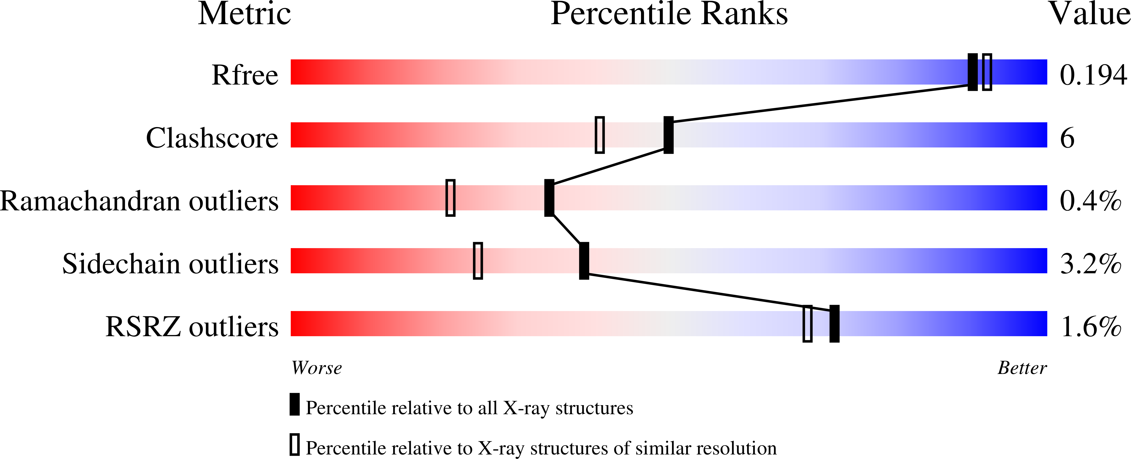
Structure analysis Details
Assembly composition:
monomeric (preferred)
Assembly name:
Carbonic anhydrase 2 (preferred)
PDBe Complex ID:
PDB-CPX-133681 (preferred)
Entry contents:
1 distinct polypeptide molecule
Macromolecule:
Carbonic anhydrase 2
Molecule details ›
Chain: A
Length: 260 amino acids
Theoretical weight: 29.29 KDa
Source organism: Homo sapiens
Expression system: Not provided
UniProt:
Sequence domains: Eukaryotic-type carbonic anhydrase
Structure domains: Alpha carbonic anhydrase
Length: 260 amino acids
Theoretical weight: 29.29 KDa
Source organism: Homo sapiens
Expression system: Not provided
UniProt:
- Canonical:
 P00918 (Residues: 1-260; Coverage: 100%)
Sequence domains: Eukaryotic-type carbonic anhydrase
Structure domains: Alpha carbonic anhydrase


