3wlc

X-ray diffraction
2.49Å resolution

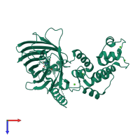
Crystal structure of dimeric GCaMP6m

Released:
Primary publication:
Structural basis of the ultrasensitive calcium indicator GCaMP6.
Sci China Life Sci 57 269-274 (2014)
PMID: 24390420

Function and Biology Details

Reaction catalysed:
ATP + [myosin light-chain] = ADP + [myosin light-chain] phosphate
Biochemical function:
Biological process:
Cellular component:
  • not assigned

Structure analysis Details

Assembly composition:
homo dimer (preferred)
PDBe Complex ID:
PDB-CPX-144018 (preferred)
Entry contents:
1 distinct polypeptide molecule
Macromolecule:
Calmodulin-1; Green fluorescent protein; Myosin light chain kinase, smooth muscle, deglutamylated form Chain: A
Molecule details ›
Chain: A
Length: 448 amino acids
Theoretical weight: 50.4 KDa
Source organisms: Expression system: Escherichia coli
UniProt:
  • Canonical: P11799 (Residues: 1731-1749, 1752-1763; Coverage: 2%)
  • Canonical: P42212 (Residues: 2-144, 191-192; Coverage: 61%)
  • Canonical: P0DP29 (Residues: 3-42, 45-149; Coverage: 97%)
Gene names: CaMI, Calm, Calm1, Cam, Cam1, GFP, Mylk
Sequence domains:
Structure domains: Green fluorescent protein

Ligands and Environments

1 bound ligand:
1 modified residue:

Experiments and Validation Details

Entry percentile scores
X-ray source: RIGAKU MICROMAX-007
Spacegroup: C2
Unit cell:
a: 124.44Å b: 46.86Å c: 68.34Å
α: 90° β: 99.36° γ: 90°
R-values:
R R work R free
0.179 0.177 0.225
Expression system: Escherichia coli