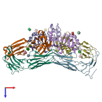
3c09

X-ray diffraction
3.2Å resolution

Crystal structure the Fab fragment of matuzumab (Fab72000) in complex with domain III of the extracellular region of EGFR

Released:

Function and Biology Details

Structure analysis Details

Assembly composition:
hetero trimer (preferred)
PDBe Complex ID:
PDB-CPX-233873 (preferred)
Entry contents:
3 distinct polypeptide molecules
Macromolecules (3 distinct):
Matuzumab Fab Light chain Chains: B, L
Molecule details ›
Chains: B, L
Length: 212 amino acids
Theoretical weight: 23.24 KDa
Source organism: Mus musculus
Expression system: Mus musculus
Structure domains: Immunoglobulins
Matuzumab Fab Heavy chain Chains: C, H
Molecule details ›
Chains: C, H
Length: 223 amino acids
Theoretical weight: 24.04 KDa
Source organism: Mus musculus
Expression system: Mus musculus
Structure domains: Immunoglobulins
Epidermal growth factor receptor Chains: A, D
Molecule details ›
Chains: A, D
Length: 214 amino acids
Theoretical weight: 23.79 KDa
Source organism: Homo sapiens
Expression system: Spodoptera frugiperda
UniProt:
  • Canonical: P00533 (Residues: 336-538; Coverage: 17%)
Gene names: EGFR, ERBB, ERBB1, HER1
Sequence domains: Receptor L domain
Structure domains: Receptor L-domain

Ligands and Environments

3 bound ligands:
No modified residues

Experiments and Validation Details

Entry percentile scores
X-ray source: SLS BEAMLINE X06SA
Spacegroup: C2
Unit cell:
a: 141.073Å b: 205.035Å c: 81.577Å
α: 90° β: 117.49° γ: 90°
R-values:
R R work R free
0.245 0.242 0.299
Expression systems:
  • Mus musculus
  • Spodoptera frugiperda