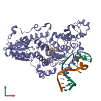
3cvy

X-ray diffraction
2.7Å resolution

Drosophila melanogaster (6-4) photolyase bound to repaired ds DNA

Released:
Source organism: Drosophila melanogaster
Primary publication:
Crystal structure and mechanism of a DNA (6-4) photolyase.
Angew Chem Int Ed Engl 47 10076-80 (2008)
PMID: 18956392

Function and Biology Details

Structure analysis Details

Assembly composition:
hetero trimer (preferred)
PDBe Complex ID:
PDB-CPX-116159 (preferred)
Entry contents:
1 distinct polypeptide molecule
2 distinct DNA molecules
Macromolecules (3 distinct):
Photolyase/cryptochrome alpha/beta domain-containing protein Chain: A
Molecule details ›
Chain: A
Length: 543 amino acids
Theoretical weight: 62.91 KDa
Source organism: Drosophila melanogaster
Expression system: Escherichia coli
UniProt:
  • Canonical: Q8SXK5 (Residues: 1-520; Coverage: 96%)
Gene names: CG2488, phr6-4
Sequence domains:
Structure domains:
DNA (5'-D(*DAP*DCP*DAP*DGP*DCP*DGP*DGP*DTP*DTP*DGP*DCP*DAP*DGP*DGP*DT)-3') Chain: C
Molecule details ›
Chain: C
Length: 15 nucleotides
Theoretical weight: 4.65 KDa
Source organism: Drosophila melanogaster
Expression system: Not provided
DNA (5'-D(*DTP*DAP*DCP*DCP*DTP*DGP*DCP*DAP*DAP*DCP*DCP*DGP*DCP*DTP*DG)-3') Chain: D
Molecule details ›
Chain: D
Length: 15 nucleotides
Theoretical weight: 4.53 KDa
Source organism: Drosophila melanogaster
Expression system: Not provided

Ligands and Environments

1 bound ligand:
No modified residues

Experiments and Validation Details

Entry percentile scores
X-ray source: SLS BEAMLINE X10SA
Spacegroup: P212121
Unit cell:
a: 83.36Å b: 88.84Å c: 86.59Å
α: 90° β: 90° γ: 90°
R-values:
R R work R free
0.215 0.212 0.266
Expression systems:
  • Escherichia coli
  • Not provided