3dih

X-ray diffraction
2.6Å resolution

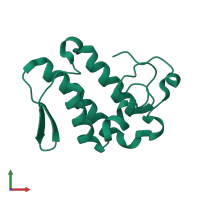
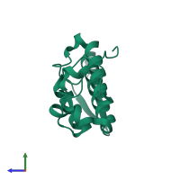
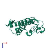
Crystal structure of ammodytin L

Released:
Source organism: Vipera ammodytes ammodytes
Entry authors: Turk D, Guncar G, Krizaj I

Function and Biology Details

Structure analysis Details

Assemblies composition:
monomeric (preferred)
homo dimer
PDBe Complex ID:
PDB-CPX-148218 (preferred)
Entry contents:
1 distinct polypeptide molecule
Macromolecule:
Basic phospholipase A2 homolog ammodytin L Chain: A
Molecule details ›
Chain: A
Length: 122 amino acids
Theoretical weight: 13.9 KDa
Source organism: Vipera ammodytes ammodytes
UniProt:
  • Canonical: P17935 (Residues: 17-138; Coverage: 100%)
Sequence domains: Phospholipase A2
Structure domains: Phospholipase A2 domain

Ligands and Environments

No bound ligands
No modified residues

Experiments and Validation Details

Entry percentile scores
X-ray source: RIGAKU RU200
Spacegroup: R32
Unit cell:
a: 120.5Å b: 120.5Å c: 63.15Å
α: 90° β: 90° γ: 120°
R-values:
R R work R free
0.159 0.156 0.226