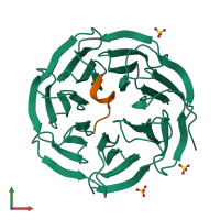
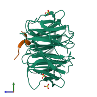
3eg6

X-ray diffraction
1.72Å resolution

Structure of WDR5 bound to MLL1 peptide

Released:
Source organism: Homo sapiens
Primary publication:
Structure of WDR5 bound to mixed lineage leukemia protein-1 peptide.
J Biol Chem 283 32158-61 (2008)
PMID: 18829459

Function and Biology Details

Reaction catalysed:
S-adenosyl-L-methionine + a [histone H3]-L-lysine(4) = S-adenosyl-L-homocysteine + a [histone H3]-N(6)-methyl-L-lysine(4)
Cellular component:

Structure analysis Details

Assembly composition:
hetero dimer (preferred)
PDBe Complex ID:
PDB-CPX-158494 (preferred)
Entry contents:
2 distinct polypeptide molecules
Macromolecules (2 distinct):
WD repeat-containing protein 5 Chain: A
Molecule details ›
Chain: A
Length: 312 amino acids
Theoretical weight: 34.3 KDa
Source organism: Homo sapiens
Expression system: Escherichia coli
UniProt:
  • Canonical: P61964 (Residues: 23-334; Coverage: 93%)
Gene names: BIG3, WDR5
Sequence domains: WD domain, G-beta repeat
Structure domains: YVTN repeat-like/Quinoprotein amine dehydrogenase
MLL cleavage product C180 Chain: C
Molecule details ›
Chain: C
Length: 13 amino acids
Theoretical weight: 1.34 KDa
Source organism: Homo sapiens
Expression system: Not provided
UniProt:
  • Canonical: Q03164 (Residues: 3762-3773; Coverage: 0%)
Gene names: ALL1, CXXC7, HRX, HTRX, KMT2A, MLL, MLL1, TRX1

Ligands and Environments

1 bound ligand:
No modified residues

Experiments and Validation Details

Entry percentile scores
X-ray source: NSLS BEAMLINE X6A
Spacegroup: C2221
Unit cell:
a: 78.254Å b: 98.384Å c: 80.075Å
α: 90° β: 90° γ: 90°
R-values:
R R work R free
0.203 0.203 0.24
Expression systems:
  • Escherichia coli
  • Not provided