3eov

X-ray diffraction
2.6Å resolution

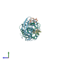
Crystal structure of cyclophilin from Leishmania donovani ligated with cyclosporin A

Released:

Function and Biology Details

Structure analysis Details

Assembly composition:
hetero dimer (preferred)
PDBe Complex ID:
PDB-CPX-163515 (preferred)
Entry contents:
2 distinct polypeptide molecules
Macromolecules (2 distinct):
Peptidyl-prolyl cis-trans isomerase Chains: A, B
Molecule details ›
Chains: A, B
Length: 172 amino acids
Theoretical weight: 19.09 KDa
Source organism: Leishmania donovani
Expression system: Escherichia coli
UniProt:
  • Canonical: Q9U9R3 (Residues: 22-187; Coverage: 100%)
Gene name: CYP
Sequence domains: Cyclophilin type peptidyl-prolyl cis-trans isomerase/CLD
Structure domains: Cyclophilin-like
CYCLOSPORIN A Chains: C, D
Molecule details ›
Chains: C, D
Length: 11 amino acids
Theoretical weight: 1.22 KDa
Source organism: Tolypocladium inflatum
Expression system: Not provided

Ligands and Environments

No bound ligands

Experiments and Validation Details

Entry percentile scores
X-ray source: RIGAKU RU200
Spacegroup: C2
Unit cell:
a: 69.81Å b: 83.125Å c: 73.58Å
α: 90° β: 103.03° γ: 90°
R-values:
R R work R free
0.207 0.207 0.251
Expression systems:
  • Escherichia coli
  • Not provided