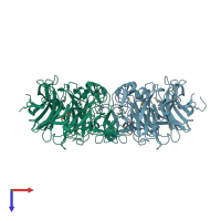
3h71

X-ray diffraction
1.7Å resolution

Crystal structure of Streptococcus pneumoniae D39 neuraminidase A precursor (NanA)

Released:
Source organism: Streptococcus pneumoniae R6
Primary publication:
Crystal structures of respiratory pathogen neuraminidases.
Biochem Biophys Res Commun 380 467-71 (2009)
PMID: 19284989

Function and Biology Details

Reaction catalysed:
Hydrolysis of alpha-(2->3)-, alpha-(2->6)-, alpha-(2->8)- glycosidic linkages of terminal sialic acid residues in oligosaccharides, glycoproteins, glycolipids, colominic acid and synthetic substrates.
Biochemical function:
Biological process:
  • not assigned
Cellular component:
  • not assigned

Structure analysis Details

Assembly composition:
monomeric (preferred)
Assembly name:
PDBe Complex ID:
PDB-CPX-158649 (preferred)
Entry contents:
1 distinct polypeptide molecule
Macromolecule:
Sialidase A Chains: A, B
Molecule details ›
Chains: A, B
Length: 477 amino acids
Theoretical weight: 54.13 KDa
Source organism: Streptococcus pneumoniae R6
Expression system: Escherichia coli
UniProt:
  • Canonical: P62576 (Residues: 317-793; Coverage: 49%)
Gene names: nanA, spr1536
Sequence domains: BNR/Asp-box repeat
Structure domains:

Ligands and Environments

No bound ligands
1 modified residue:

Experiments and Validation Details

Entry percentile scores
X-ray source: NSLS BEAMLINE X4A
Spacegroup: C2
Unit cell:
a: 158.04Å b: 47.67Å c: 137.27Å
α: 90° β: 116.56° γ: 90°
R-values:
R R work R free
0.172 0.172 0.198
Expression system: Escherichia coli