3j32

Electron Microscopy
4.5Å resolution

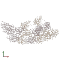
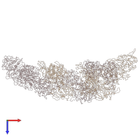
An asymmetric unit map from electron cryo-microscopy of Haliotis diversicolor molluscan hemocyanin isoform 1 (HdH1)

Released:

Function and Biology Details

Structure analysis Details

Assembly composition:
homo icosamer (preferred)
PDBe Complex ID:
PDB-CPX-111699 (preferred)
Entry contents:
1 distinct polypeptide molecule
Macromolecule:
Tyrosinase copper-binding domain-containing protein Chains: A, B
Molecule details ›
Chains: A, B
Length: 3303 amino acids
Theoretical weight: 378.67 KDa
Source organism: Haliotis diversicolor
UniProt:
  • Canonical: C7FEG7 (Residues: 1-3235; Coverage: 97%)
Gene name: H1
Sequence domains:

Ligands and Environments

No bound ligands
No modified residues

Experiments and Validation Details

Entry percentile scores
Resolution: 4.5Å
Relevant EMDB volumes: EMD-5586