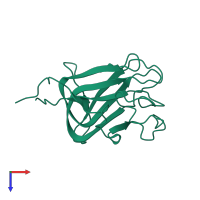
3jd6

Electron Microscopy
4.1Å resolution

Double octamer structure of retinoschisin, a cell-cell adhesion protein of the retina

Released:
Source organism: Homo sapiens
Related structures: EMD-6425

Function and Biology Details

Structure analysis Details

Assembly composition:
homo hexadecamer (preferred)
Assembly name:
PDBe Complex ID:
PDB-CPX-127522 (preferred)
Entry contents:
1 distinct polypeptide molecule
Macromolecule:
Retinoschisin Chain: O
Molecule details ›
Chain: O
Length: 207 amino acids
Theoretical weight: 23.89 KDa
Source organism: Homo sapiens
Expression system: Spodoptera frugiperda
UniProt:
  • Canonical: O15537 (Residues: 24-224; Coverage: 100%)
Gene names: RS1, XLRS1
Sequence domains: F5/8 type C domain

Ligands and Environments

No bound ligands
No modified residues

Experiments and Validation Details

Entry percentile scores
Resolution: 4.1Å
Relevant EMDB volumes: EMD-6425
Expression system: Spodoptera frugiperda