3lk7

X-ray diffraction
1.5Å resolution

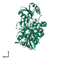
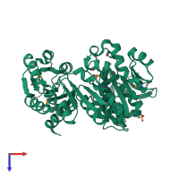
The Crystal Structure of UDP-N-acetylmuramoylalanine-D-glutamate (MurD) ligase from Streptococcus agalactiae to 1.5A

Released:
Entry authors: Stein AJ, Sather A, Shakelford G, Joachimiak A, Midwest Center for Structural Genomics (MCSG)

Function and Biology Details

Reaction catalysed:
ATP + UDP-N-acetyl-alpha-D-muramoyl-L-alanine + D-glutamate = ADP + phosphate + UDP-N-acetyl-alpha-D-muramoyl-L-alanyl-D-glutamate
Biochemical function:
Biological process:
Cellular component:

Structure analysis Details

Assembly composition:
monomeric (preferred)
PDBe Complex ID:
PDB-CPX-184290 (preferred)
Entry contents:
1 distinct polypeptide molecule
Macromolecule:
UDP-N-acetylmuramoylalanine--D-glutamate ligase Chain: A
Molecule details ›
Chain: A
Length: 451 amino acids
Theoretical weight: 49.5 KDa
Source organism: Streptococcus agalactiae serogroup V
Expression system: Escherichia coli BL21(DE3)
UniProt:
  • Canonical: Q8E186 (Residues: 1-451; Coverage: 100%)
Gene names: SAG0475, murD
Sequence domains:
Structure domains:

Ligands and Environments

2 bound ligands:
1 modified residue:

Experiments and Validation Details

Entry percentile scores
X-ray source: APS BEAMLINE 19-ID
Spacegroup: C2
Unit cell:
a: 161.539Å b: 65.045Å c: 52.889Å
α: 90° β: 107.52° γ: 90°
R-values:
R R work R free
0.188 0.187 0.208
Expression system: Escherichia coli BL21(DE3)