Function and Biology Details
Biochemical function:
Biological process:
Cellular component:
- not assigned
Sequence domains:
- Glycoside hydrolase family 38, central domain
- Glycosyl hydrolases family 38, C-terminal beta sandwich domain
- Glycosyl hydrolase family 38, C-terminal
- Glycosyl hydrolases 38, beta-1 domain
- Galactose mutarotase-like domain superfamily
- Glycoside hydrolase families 57/38, central domain superfamily
- Glycoside hydrolase family 38, central domain superfamily
- Glycoside hydrolase family 38, N-terminal domain
2 more domains
Structure analysis Details
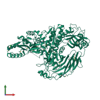
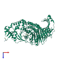
Assemblies composition:
Assembly name:
PDBe Complex ID:
PDB-CPX-182795 (preferred)
Entry contents:
1 distinct polypeptide molecule
Macromolecule:


