Function and Biology Details
Reaction catalysed:
Methyl-CoM + CoB = CoM-S-S-CoB + methane
Biochemical function:
Biological process:
Cellular component:
Sequence domains:
- Methyl-coenzyme M reductase, gamma subunit
- Methyl-coenzyme M reductase, alpha/beta subunit, C-terminal
- Methyl-coenzyme M reductase, ferredoxin-like fold
- Methyl-coenzyme M reductase, beta subunit
- Methyl-coenzyme M reductase, alpha subunit, N-terminal subdomain 2
- Methyl coenzyme M reductase, alpha subunit
- Methyl-coenzyme M reductase, gamma subunit superfamily
- Methyl-coenzyme M reductase, beta subunit, C-terminal
4 more domains
Structure analysis Details
Assembly composition:
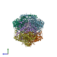
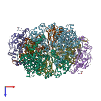
hetero hexamer (preferred)
Assembly name:
Methyl-coenzyme M Reductase (preferred)
PDBe Complex ID:
PDB-CPX-145876 (preferred)
Entry contents:
3 distinct polypeptide molecules
Macromolecules (3 distinct):
Ligands and Environments
Experiments and Validation Details
wwPDB Validation report is not available for this entry.
X-ray source:
APS BEAMLINE 14-BM-C
Spacegroup:
P21


