3neg

X-ray diffraction
2.8Å resolution

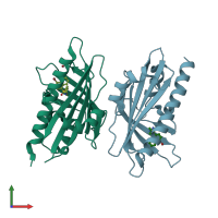
Pyrabactin-bound PYL1 structure in the open and close forms

Released:
Source organism: Arabidopsis thaliana
Primary publication:
Functional mechanism of the abscisic acid agonist pyrabactin.
J Biol Chem 285 28946-52 (2010)
PMID: 20554531

Function and Biology Details

Reaction catalysed:
[a protein]-serine/threonine phosphate + H(2)O = [a protein]-serine/threonine + phosphate

Structure analysis Details

Assembly composition:
monomeric (preferred)
PDBe Complex ID:
PDB-CPX-186652 (preferred)
Entry contents:
1 distinct polypeptide molecule
Macromolecule:
Abscisic acid receptor PYL1 Chains: A, B
Molecule details ›
Chains: A, B
Length: 202 amino acids
Theoretical weight: 23.33 KDa
Source organism: Arabidopsis thaliana
Expression system: Escherichia coli BL21(DE3)
UniProt:
  • Canonical: Q8VZS8 (Residues: 20-221; Coverage: 91%)
Gene names: At5g46790, MZA15.21, PYL1, RCAR12
Sequence domains: Polyketide cyclase / dehydrase and lipid transport
Structure domains: Alpha-D-Glucose-1,6-Bisphosphate; Chain A, domain 4

Ligands and Environments

2 bound ligands:
No modified residues

Experiments and Validation Details

Entry percentile scores
X-ray source: SSRF BEAMLINE BL17U
Spacegroup: P3121
Unit cell:
a: 130.23Å b: 130.23Å c: 88.25Å
α: 90° β: 90° γ: 120°
R-values:
R R work R free
0.197 0.195 0.242
Expression system: Escherichia coli BL21(DE3)