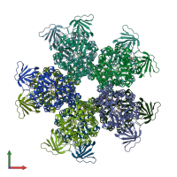
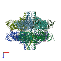
3ooq

X-ray diffraction
2.06Å resolution

CRYSTAL STRUCTURE OF amidohydrolase from Thermotoga maritima MSB8

Released:
Source organism: Thermotoga maritima MSB8
Entry authors: Malashkevich VN, Toro R, Sauder JM, Burley SK, Almo SC, New York SGX Research Center for Structural Genomics (NYSGXRC)

Function and Biology Details

Structure analysis Details

Assembly composition:
homo decamer (preferred)
PDBe Complex ID:
PDB-CPX-194863 (preferred)
Entry contents:
1 distinct polypeptide molecule
Macromolecule:
Amidohydrolase-related domain-containing protein Chains: A, B, C, D, E, F, G, H, I, J
Molecule details ›
Chains: A, B, C, D, E, F, G, H, I, J
Length: 396 amino acids
Theoretical weight: 44.38 KDa
Source organism: Thermotoga maritima MSB8
Expression system: Escherichia coli BL21(DE3)
UniProt:
  • Canonical: Q9X247 (Residues: 2-386; Coverage: 100%)
Gene name: TM_1723
Sequence domains: Amidohydrolase family
Structure domains:

Ligands and Environments

1 bound ligand:
1 modified residue:

Experiments and Validation Details

Entry percentile scores
X-ray source: NSLS BEAMLINE X29A
Spacegroup: C2
Unit cell:
a: 152.45Å b: 148.3Å c: 195.02Å
α: 90° β: 105.31° γ: 90°
R-values:
R R work R free
0.17 0.167 0.213
Expression system: Escherichia coli BL21(DE3)