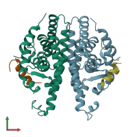
3r29

X-ray diffraction
2.9Å resolution

Crystal structure of RXRalpha ligand-binding domain complexed with corepressor SMRT2

Released:
Source organism: Homo sapiens
Primary publication:
Structural basis for retinoic X receptor repression on the tetramer.
J Biol Chem 286 24593-8 (2011)
PMID: 21613212

Function and Biology Details

Structure analysis Details

Assembly composition:
hetero octamer (preferred)
PDBe Complex ID:
PDB-CPX-148732 (preferred)
Entry contents:
2 distinct polypeptide molecules
Macromolecules (2 distinct):
Retinoic acid receptor RXR-alpha Chains: A, B
Molecule details ›
Chains: A, B
Length: 240 amino acids
Theoretical weight: 26.86 KDa
Source organism: Homo sapiens
Expression system: Escherichia coli
UniProt:
  • Canonical: P19793 (Residues: 223-462; Coverage: 52%)
Gene names: NR2B1, RXRA
Sequence domains: Ligand-binding domain of nuclear hormone receptor
Structure domains: Retinoid X Receptor
Nuclear receptor corepressor 2 Chains: C, D
Molecule details ›
Chains: C, D
Length: 16 amino acids
Theoretical weight: 1.75 KDa
Source organism: Homo sapiens
Expression system: Not provided
UniProt:
  • Canonical: Q9Y618 (Residues: 2335-2350; Coverage: 1%)
Gene names: CTG26, NCOR2

Ligands and Environments

No bound ligands
No modified residues

Experiments and Validation Details

Entry percentile scores
X-ray source: SSRF BEAMLINE BL17U
Spacegroup: P41212
Unit cell:
a: 118.22Å b: 118.22Å c: 84.02Å
α: 90° β: 90° γ: 90°
R-values:
R R work R free
0.244 0.24 0.326
Expression systems:
  • Escherichia coli
  • Not provided