3s04

X-ray diffraction
2.44Å resolution

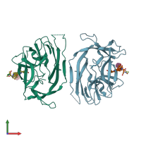
Crystal structure of Escherichia coli type I signal peptidase in complex with an Arylomycin Lipoglycopeptide Antibiotic

Released:

Function and Biology Details

Structure analysis Details

Assemblies composition:
hetero dimer (preferred)
hetero tetramer
PDBe Complex ID:
PDB-CPX-133587 (preferred)
Entry contents:
2 distinct polypeptide molecules
Macromolecules (2 distinct):
Signal peptidase I Chains: A, B
Molecule details ›
Chains: A, B
Length: 250 amino acids
Theoretical weight: 28.08 KDa
Source organism: Escherichia coli K-12
Expression system: Escherichia coli BL21(DE3)
UniProt:
  • Canonical: P00803 (Residues: 75-324; Coverage: 77%)
Gene names: JW2552, b2568, lepB
Sequence domains: Signal peptidase, peptidase S26
Structure domains:
Glyco-Arylomycin Chains: I, J
Molecule details ›
Chains: I, J
Length: 6 amino acids
Theoretical weight: 661 Da
Source organism: Streptomyces sp.

Ligands and Environments

2 bound ligands:
2 modified residues:

Experiments and Validation Details

Entry percentile scores
X-ray source: RIGAKU MICROMAX-007
Spacegroup: P43212
Unit cell:
a: 72.01Å b: 72.01Å c: 262.57Å
α: 90° β: 90° γ: 90°
R-values:
R R work R free
0.246 0.245 0.265
Expression system: Escherichia coli BL21(DE3)