3t1u

X-ray diffraction
2Å resolution

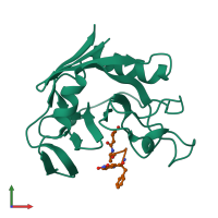
Crystal Structure of the complex of Cyclophilin-A enzyme from Azotobacter vinelandii with sucAFPFpNA peptide

Released:
Source organism: Azotobacter vinelandii DJ
Primary publication:
Structure of a bacterial cytoplasmic cyclophilin A in complex with a tetrapeptide.
Acta Crystallogr Sect F Struct Biol Cryst Commun 68 259-64 (2012)
PMID: 22442217

Function and Biology Details

Structure analysis Details

Assembly composition:
hetero dimer (preferred)
PDBe Complex ID:
PDB-CPX-111090 (preferred)
Entry contents:
2 distinct polypeptide molecules
Macromolecules (2 distinct):
Peptidyl-prolyl cis-trans isomerase Chain: A
Molecule details ›
Chain: A
Length: 163 amino acids
Theoretical weight: 17.68 KDa
Source organism: Azotobacter vinelandii DJ
Expression system: Escherichia coli BL21(DE3)
UniProt:
  • Canonical: C1DHE4 (Residues: 2-163; Coverage: 99%)
Gene name: Avin_23510
Sequence domains: Cyclophilin type peptidyl-prolyl cis-trans isomerase/CLD
Structure domains: Cyclophilin-like
succinyl-Ala-Phe-Pro-Phe-p-nitroanilide Chain: B
Molecule details ›
Chain: B
Length: 6 amino acids
Theoretical weight: 701 Da
Source organism: Azotobacter vinelandii DJ
Expression system: Not provided

Ligands and Environments

No bound ligands
No modified residues

Experiments and Validation Details

Entry percentile scores
X-ray source: RIGAKU
Spacegroup: P42212
Unit cell:
a: 66.41Å b: 66.41Å c: 71.53Å
α: 90° β: 90° γ: 90°
R-values:
R R work R free
0.201 0.201 0.238
Expression systems:
  • Escherichia coli BL21(DE3)
  • Not provided