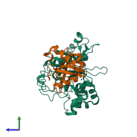
3vh3

X-ray diffraction
2Å resolution

Crystal structure of Atg7CTD-Atg8 complex

Released:

Function and Biology Details

Structure analysis Details

Assembly composition:
hetero tetramer (preferred)
PDBe Complex ID:
PDB-CPX-153573 (preferred)
Entry contents:
2 distinct polypeptide molecules
Macromolecules (2 distinct):
Ubiquitin-like modifier-activating enzyme ATG7 Chain: A
Molecule details ›
Chain: A
Length: 340 amino acids
Theoretical weight: 38.15 KDa
Source organism: Saccharomyces cerevisiae S288C
Expression system: Escherichia coli
UniProt:
  • Canonical: P38862 (Residues: 295-630; Coverage: 53%)
Gene names: APG7, ATG7, CVT2, YHR171W
Sequence domains: ThiF family
Structure domains: NAD(P)-binding Rossmann-like Domain
Autophagy-related protein 8 Chain: B
Molecule details ›
Chain: B
Length: 119 amino acids
Theoretical weight: 13.75 KDa
Source organism: Saccharomyces cerevisiae S288C
Expression system: Escherichia coli
UniProt:
  • Canonical: P38182 (Residues: 1-116; Coverage: 99%)
Gene names: APG8, ATG8, AUT7, CVT5, YBL0732, YBL078C
Sequence domains: Autophagy protein Atg8 ubiquitin like
Structure domains: Phosphatidylinositol 3-kinase Catalytic Subunit; Chain A, domain 1

Ligands and Environments

1 bound ligand:
No modified residues

Experiments and Validation Details

Entry percentile scores
X-ray source: SPRING-8 BEAMLINE BL41XU
Spacegroup: P43212
Unit cell:
a: 69.815Å b: 69.815Å c: 220.392Å
α: 90° β: 90° γ: 90°
R-values:
R R work R free
0.235 0.232 0.257
Expression system: Escherichia coli