4h6s

X-ray diffraction
2.19Å resolution

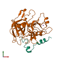
Crystal structure of thrombin mutant E14eA/D14lA/E18A/S195A

Released:
Source organism: Homo sapiens
Primary publication:
Autoactivation of thrombin precursors.
J Biol Chem 288 11601-10 (2013)
PMID: 23467412

Function and Biology Details

Structure analysis Details

Assembly composition:
hetero dimer (preferred)
Assembly name:
PDBe Complex ID:
PDB-CPX-133055 (preferred)
Entry contents:
2 distinct polypeptide molecules
Macromolecules (2 distinct):
Thrombin light chain Chain: A
Molecule details ›
Chain: A
Length: 31 amino acids
Theoretical weight: 3.55 KDa
Source organism: Homo sapiens
Expression system: Escherichia coli
UniProt:
  • Canonical: P00734 (Residues: 333-363; Coverage: 5%)
Gene name: F2
Sequence domains: Thrombin light chain
Thrombin heavy chain Chain: B
Molecule details ›
Chain: B
Length: 259 amino acids
Theoretical weight: 29.71 KDa
Source organism: Homo sapiens
Expression system: Escherichia coli
UniProt:
  • Canonical: P00734 (Residues: 364-622; Coverage: 43%)
Gene name: F2
Sequence domains: Trypsin
Structure domains: Trypsin-like serine proteases

Ligands and Environments

1 bound ligand:
No modified residues

Experiments and Validation Details

Entry percentile scores
X-ray source: RIGAKU MICROMAX-007 HF
Spacegroup: P21212
Unit cell:
a: 63.904Å b: 70.828Å c: 56.103Å
α: 90° β: 90° γ: 90°
R-values:
R R work R free
0.199 0.196 0.259
Expression system: Escherichia coli