


Function and Biology Details
Reaction catalysed:
Strict requirement for an Asp residue at position P1 and has a preferred cleavage sequence of Asp-Glu-Val-Asp-|-
Biochemical function:
Biological process:
Cellular component:
- not assigned
Structure analysis Details
Assembly composition:
hetero tetramer (preferred)
Assembly name:
Caspase-7 and peptide (preferred)
PDBe Complex ID:
PDB-CPX-157259 (preferred)
Entry contents:
2 distinct polypeptide molecules
Macromolecules (2 distinct):
Caspase-7 subunit p20
Molecule details ›
Chains: A, B
Length: 272 amino acids
Theoretical weight: 31.01 KDa
Source organism: Homo sapiens
Expression system: Escherichia coli
UniProt:
Sequence domains: Caspase domain
Structure domains: Rossmann fold
Length: 272 amino acids
Theoretical weight: 31.01 KDa
Source organism: Homo sapiens
Expression system: Escherichia coli
UniProt:
- Canonical:
 P55210 (Residues: 47-204, 205-303; Coverage: 85%)
Sequence domains: Caspase domain
Structure domains: Rossmann fold
Ligands and Environments
No bound ligands
No modified residues
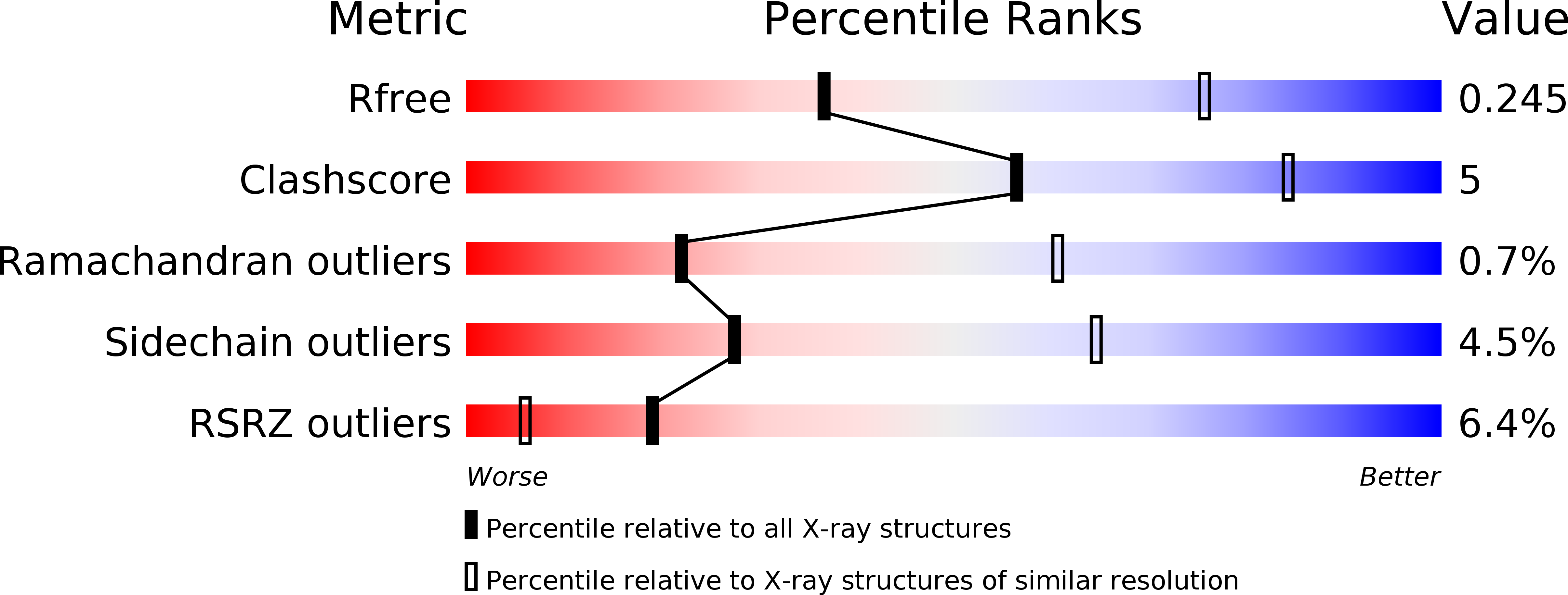
Experiments and Validation Details

X-ray source:
PAL/PLS BEAMLINE 6C1
Spacegroup:
P3221
Expression system: Escherichia coli