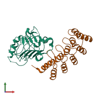
4hrl

X-ray diffraction
2.55Å resolution

Structural Basis for Eliciting a Cytotoxic Effect in HER2-Overexpressing Cancer Cells via Binding to the Extracellular Domain of HER2

Released:

Function and Biology Details

Reaction catalysed:
ATP + a [protein]-L-tyrosine = ADP + a [protein]-L-tyrosine phosphate
Biochemical function:
  • not assigned
Biological process:
  • not assigned
Cellular component:
  • not assigned

Structure analysis Details

Assembly composition:
hetero dimer (preferred)
PDBe Complex ID:
PDB-CPX-138066 (preferred)
Entry contents:
2 distinct polypeptide molecules
Macromolecules (2 distinct):
Receptor tyrosine-protein kinase erbB-2 Chain: C
Molecule details ›
Chain: C
Length: 198 amino acids
Theoretical weight: 22.21 KDa
Source organism: Homo sapiens
Expression system: Spodoptera frugiperda
UniProt:
  • Canonical: P04626 (Residues: 24-219; Coverage: 16%)
Gene names: ERBB2, HER2, MLN19, NEU, NGL
Sequence domains: Receptor L domain
Structure domains: Receptor L-domain
Designed Ankyrin Repeat Protein 9_29 Chain: A
Molecule details ›
Chain: A
Length: 171 amino acids
Theoretical weight: 18.56 KDa
Source organism: synthetic construct
Expression system: Escherichia coli
Structure domains: Ankyrin repeat-containing domain

Ligands and Environments

No bound ligands
No modified residues

Experiments and Validation Details

Entry percentile scores
X-ray source: SLS BEAMLINE X06SA
Spacegroup: P212121
Unit cell:
a: 46.6Å b: 80.5Å c: 115.1Å
α: 90° β: 90° γ: 90°
R-values:
R R work R free
0.203 0.2 0.253
Expression systems:
  • Spodoptera frugiperda
  • Escherichia coli