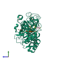
4kmd

X-ray diffraction
1.7Å resolution

Crystal structure of Sufud60-Gli1p

Released:

Function and Biology Details

Structure analysis Details

Assemblies composition:
hetero dimer (preferred)
hetero tetramer
Assembly name:
PDBe Complex ID:
PDB-CPX-139936 (preferred)
Entry contents:
2 distinct polypeptide molecules
Macromolecules (2 distinct):
Suppressor of fused homolog Chain: A
Molecule details ›
Chain: A
Length: 444 amino acids
Theoretical weight: 49.38 KDa
Source organism: Homo sapiens
Expression system: Escherichia coli
UniProt:
  • Canonical: Q9UMX1 (Residues: 1-484; Coverage: 88%)
Gene names: SUFU, UNQ650/PRO1280
Sequence domains:
Structure domains: Sufu, C-terminal domain
Zinc finger protein GLI1 Chain: B
Molecule details ›
Chain: B
Length: 17 amino acids
Theoretical weight: 1.68 KDa
Source organism: Homo sapiens
Expression system: Not provided
UniProt:
  • Canonical: P08151 (Residues: 112-128; Coverage: 2%)
Gene names: GLI, GLI1

Ligands and Environments

2 bound ligands:
No modified residues

Experiments and Validation Details

Entry percentile scores
X-ray source: SSRF BEAMLINE BL17U
Spacegroup: C2221
Unit cell:
a: 70.83Å b: 82.118Å c: 149.756Å
α: 90° β: 90° γ: 90°
R-values:
R R work R free
0.196 0.195 0.222
Expression systems:
  • Escherichia coli
  • Not provided