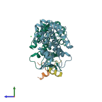
4oyk

X-ray diffraction
2Å resolution

Structure of HOIP PUB domain bound to OTULIN PIM

Released:

Function and Biology Details

Reactions catalysed:
(1a) [E2 ubiquitin-conjugating enzyme]-S-ubiquitinyl-L-cysteine + [RBR-type E3 ubiquitin transferase]-L-cysteine = [E2 ubiquitin-conjugating enzyme]-L-cysteine + [RBR-type E3 ubiquitin transferase]-S-ubiquitinyl-L-cysteine
Thiol-dependent hydrolysis of ester, thioester, amide, peptide and isopeptide bonds formed by the C-terminal Gly of ubiquitin (a 76-residue protein attached to proteins as an intracellular targeting signal).
Biological process:
  • not assigned
Cellular component:

Structure analysis Details

Assembly composition:
hetero dimer (preferred)
PDBe Complex ID:
PDB-CPX-188379 (preferred)
Entry contents:
2 distinct polypeptide molecules
Macromolecules (2 distinct):
E3 ubiquitin-protein ligase RNF31 Chains: A, B
Molecule details ›
Chains: A, B
Length: 177 amino acids
Theoretical weight: 20.1 KDa
Source organism: Homo sapiens
Expression system: Escherichia coli BL21(DE3)
UniProt:
  • Canonical: Q96EP0 (Residues: 3-179; Coverage: 17%)
Gene names: RNF31, ZIBRA
Sequence domains:
Structure domains: Methane Monooxygenase Hydroxylase; Chain G, domain 1
Ubiquitin thioesterase otulin Chains: C, D
Molecule details ›
Chains: C, D
Length: 19 amino acids
Theoretical weight: 2.33 KDa
Source organism: Homo sapiens
Expression system: Not provided
UniProt:
  • Canonical: Q96BN8 (Residues: 49-67; Coverage: 5%)
Gene names: FAM105B, OTULIN

Ligands and Environments

1 bound ligand:
No modified residues

Experiments and Validation Details

Entry percentile scores
X-ray source: DIAMOND BEAMLINE I02
Spacegroup: P61
Unit cell:
a: 64.049Å b: 64.049Å c: 172.022Å
α: 90° β: 90° γ: 120°
R-values:
R R work R free
0.202 0.2 0.237
Expression systems:
  • Escherichia coli BL21(DE3)
  • Not provided