Function and Biology Details
Reaction catalysed:
1-phosphatidyl-1D-myo-inositol 4,5-bisphosphate + H(2)O = 1D-myo-inositol 1,4,5-trisphosphate + diacylglycerol
Biochemical function:
Biological process:
Cellular component:
Sequence domains:
- Guanine nucleotide binding protein (G-protein), alpha subunit
- Phosphoinositide phospholipase C family
- G-protein alpha subunit, group Q
- C2 domain
- C2 domain superfamily
- Phospholipase C, phosphatidylinositol-specific, Y domain
- Phosphatidylinositol-specific phospholipase C, X domain
- PLC-like phosphodiesterase, TIM beta/alpha-barrel domain superfamily
5 more domains
Structure analysis Details
Assembly composition:
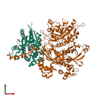
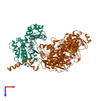
hetero dimer (preferred)
Assembly name:
PDBe Complex ID:
PDB-CPX-149200 (preferred)
Entry contents:
2 distinct polypeptide molecules
Macromolecules (2 distinct):


