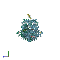
4qlj

X-ray diffraction
1.95Å resolution

Crystal structure of rice BGlu1 E386G/Y341A/Q187A mutant complexed with cellotetraose

Released:

Function and Biology Details

Reaction catalysed:
Hydrolysis of terminal, non-reducing beta-D-glucosyl residues with release of beta-D-glucose
Biochemical function:
Biological process:
Cellular component:

Structure analysis Details

Assembly composition:
monomeric (preferred)
Assembly name:
PDBe Complex ID:
PDB-CPX-181114 (preferred)
Entry contents:
1 distinct polypeptide molecule
Macromolecules (2 distinct):
Beta-glucosidase 7 Chains: A, B
Molecule details ›
Chains: A, B
Length: 481 amino acids
Theoretical weight: 54.51 KDa
Source organism: Oryza sativa Japonica Group
Expression system: Escherichia coli
UniProt:
  • Canonical: Q75I93 (Residues: 29-504; Coverage: 100%)
Gene names: BGLU1, BGLU7, LOC_Os03g49600, OSJNBa0004L11.16, Os03g0703000
Sequence domains: Glycosyl hydrolase family 1
Structure domains: Glycosidases

Ligands and Environments

Carbohydrate polymer : NEW Components: BGC
3 bound ligands:
No modified residues

Experiments and Validation Details

Entry percentile scores
X-ray source: NSRRC BEAMLINE BL13B1
Spacegroup: P212121
Unit cell:
a: 79.505Å b: 101.239Å c: 127.349Å
α: 90° β: 90° γ: 90°
R-values:
R R work R free
0.175 0.173 0.202
Expression system: Escherichia coli