4r9d

X-ray diffraction
1.24Å resolution

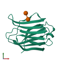
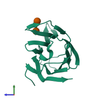
Crystal structure of Human galectin-3 CRD in complex with lactose (pH 7.9, PEG6000)

Released:
Source organism: Homo sapiens
Primary publication:
The water network in galectin-3 ligand binding site guides inhibitor design.
Acta Biochim Biophys Sin (Shanghai) 47 192-8 (2015)
PMID: 25662390

Function and Biology Details

Biochemical function:
Biological process:
  • not assigned
Cellular component:
  • not assigned

Structure analysis Details

Assembly composition:
monomeric (preferred)
Assembly name:
PDBe Complex ID:
PDB-CPX-148215 (preferred)
Entry contents:
1 distinct polypeptide molecule
Macromolecules (2 distinct):
Galectin-3 Chain: A
Molecule details ›
Chain: A
Length: 144 amino acids
Theoretical weight: 16.24 KDa
Source organism: Homo sapiens
Expression system: Escherichia coli
UniProt:
  • Canonical: P17931 (Residues: 111-250; Coverage: 56%)
Gene names: LGALS3, MAC2
Sequence domains: Galactoside-binding lectin
Structure domains: Jelly Rolls

Ligands and Environments

Carbohydrate polymer : NEW Components: BGC, GAL
No bound ligands
No modified residues

Experiments and Validation Details

Entry percentile scores
X-ray source: SSRF BEAMLINE BL17U
Spacegroup: P212121
Unit cell:
a: 36.085Å b: 57.021Å c: 61.85Å
α: 90° β: 90° γ: 90°
R-values:
R R work R free
0.172 0.172 0.18
Expression system: Escherichia coli