4wjn

X-ray diffraction
1.5Å resolution

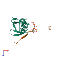
Crystal structure of SUMO1 in complex with phosphorylated PML

Released:

Function and Biology Details

Biochemical function:
  • not assigned
Biological process:
  • not assigned
Cellular component:
  • not assigned

Structure analysis Details

Assembly composition:
hetero dimer (preferred)
PDBe Complex ID:
PDB-CPX-151545 (preferred)
Entry contents:
2 distinct polypeptide molecules
Macromolecules (2 distinct):
Small ubiquitin-related modifier 1 Chain: A
Molecule details ›
Chain: A
Length: 83 amino acids
Theoretical weight: 9.53 KDa
Source organism: Homo sapiens
Expression system: Escherichia coli
UniProt:
  • Canonical: P63165 (Residues: 17-97; Coverage: 80%)
Gene names: OK/SW-cl.43, SMT3C, SMT3H3, SUMO1, UBL1
Sequence domains: Ubiquitin-2 like Rad60 SUMO-like
Structure domains: Phosphatidylinositol 3-kinase Catalytic Subunit; Chain A, domain 1
Protein PML Chain: B
Molecule details ›
Chain: B
Length: 29 amino acids
Theoretical weight: 3.3 KDa
Source organism: Homo sapiens
Expression system: Escherichia coli
UniProt:
  • Canonical: P29590 (Residues: 547-573; Coverage: 3%)
Gene names: MYL, PML, PP8675, RNF71, TRIM19

Ligands and Environments

No bound ligands
1 modified residue:

Experiments and Validation Details

Entry percentile scores
X-ray source: CLSI BEAMLINE 08ID-1
Spacegroup: P212121
Unit cell:
a: 38.274Å b: 47.079Å c: 63.986Å
α: 90° β: 90° γ: 90°
R-values:
R R work R free
0.16 0.158 0.192
Expression system: Escherichia coli