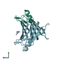
4wns

X-ray diffraction
1.4Å resolution

Crystal structure of Transthyretin complexed with pterostilbene

Released:
Source organism: Homo sapiens
Primary publication:
Structural evidence for asymmetric ligand binding to transthyretin.
Acta Crystallogr D Biol Crystallogr 71 1582-92 (2015)
PMID: 26249340

Function and Biology Details

Structure analysis Details

Assembly composition:
homo tetramer (preferred)
Assembly name:
PDBe Complex ID:
PDB-CPX-136168 (preferred)
Entry contents:
1 distinct polypeptide molecule
Macromolecule:
Transthyretin Chains: A, B
Molecule details ›
Chains: A, B
Length: 127 amino acids
Theoretical weight: 13.78 KDa
Source organism: Homo sapiens
Expression system: Escherichia coli 'BL21-Gold(DE3)pLysS AG'
UniProt:
  • Canonical: P02766 (Residues: 21-147; Coverage: 100%)
Gene names: PALB, TTR
Sequence domains: HIUase/Transthyretin family
Structure domains: Transthyretin/hydroxyisourate hydrolase domain

Ligands and Environments

3 bound ligands:
No modified residues

Experiments and Validation Details

Entry percentile scores
X-ray source: SLS BEAMLINE X06DA
Spacegroup: P21212
Unit cell:
a: 42.702Å b: 85.583Å c: 64.153Å
α: 90° β: 90° γ: 90°
R-values:
R R work R free
0.168 0.168 0.186
Expression system: Escherichia coli 'BL21-Gold(DE3)pLysS AG'