4xst

X-ray diffraction
3Å resolution

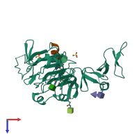
Structure of the endoglycosidase-H treated L1-CR domains of the human insulin receptor in complex with residues 697-719 of the human insulin receptor (A-isoform)

Released:

Function and Biology Details

Reaction catalysed:
ATP + a [protein]-L-tyrosine = ADP + a [protein]-L-tyrosine phosphate
Biochemical function:
  • not assigned
Biological process:
  • not assigned
Cellular component:
  • not assigned

Structure analysis Details

Assemblies composition:
hetero dimer (preferred)
hetero dodecamer
Assembly name:
PDBe Complex ID:
PDB-CPX-138933 (preferred)
Entry contents:
2 distinct polypeptide molecules
Macromolecules (3 distinct):
Insulin receptor subunit alpha Chain: E
Molecule details ›
Chain: E
Length: 317 amino acids
Theoretical weight: 36.23 KDa
Source organism: Homo sapiens
Expression system: Cricetulus griseus
UniProt:
  • Canonical: P06213 (Residues: 28-337; Coverage: 23%)
Gene name: INSR
Sequence domains:
Structure domains:
Insulin receptor subunit alpha Chain: F
Molecule details ›
Chain: F
Length: 23 amino acids
Theoretical weight: 2.79 KDa
Source organism: Rattus norvegicus
Expression system: Not provided
UniProt:
  • Canonical: P15127 (Residues: 726-748; Coverage: 2%)
Gene name: Insr

Ligands and Environments

Carbohydrate polymer : NEW Components: NAG, FUC
2 bound ligands:
No modified residues

Experiments and Validation Details

Entry percentile scores
X-ray source: AUSTRALIAN SYNCHROTRON BEAMLINE MX2
Spacegroup: P6322
Unit cell:
a: 158.718Å b: 158.718Å c: 85.912Å
α: 90° β: 90° γ: 120°
R-values:
R R work R free
0.219 0.217 0.262
Expression systems:
  • Cricetulus griseus
  • Not provided