4zlk

X-ray diffraction
2.5Å resolution

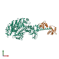
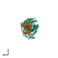
Crystal structure of mouse myosin-5a in complex with calcium-bound calmodulin

Released:
Source organism: Mus musculus
Entry authors: Shen M, Zhang N, Zheng S, Zhang W-B, Zhang H-M, Lu Z, Su QP, Sun Y, Ye K, Li X-D

Function and Biology Details

Structure analysis Details

Assembly composition:
hetero dimer (preferred)
PDBe Complex ID:
PDB-CPX-144011 (preferred)
Entry contents:
2 distinct polypeptide molecules
Macromolecules (2 distinct):
Unconventional myosin-Va Chain: A
Molecule details ›
Chain: A
Length: 839 amino acids
Theoretical weight: 96.83 KDa
Source organism: Mus musculus
Expression system: Spodoptera frugiperda
UniProt:
  • Canonical: Q99104 (Residues: 1-791; Coverage: 43%)
Gene names: Dilute, Myo5a
Sequence domains:
Calmodulin-1 Chain: B
Molecule details ›
Chain: B
Length: 149 amino acids
Theoretical weight: 16.85 KDa
Source organism: Mus musculus
Expression system: Spodoptera frugiperda
UniProt:
  • Canonical: P0DP26 (Residues: 1-149; Coverage: 100%)
Gene names: Calm, Calm1, Cam, Cam1
Sequence domains: EF-hand domain pair

Ligands and Environments

1 bound ligand:
No modified residues

Experiments and Validation Details

Entry percentile scores
X-ray source: SSRF BEAMLINE BL17U
Spacegroup: P212121
Unit cell:
a: 56.079Å b: 99.455Å c: 248.159Å
α: 90° β: 90° γ: 90°
R-values:
R R work R free
0.199 0.196 0.246
Expression system: Spodoptera frugiperda