4e54

X-ray diffraction
2.85Å resolution

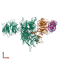
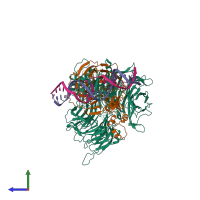
Damaged DNA induced UV-damaged DNA-binding protein (UV-DDB) dimerization and its roles in chromatinized DNA repair

Released:

Function and Biology Details

Structure analysis Details

Assembly composition:
hetero tetramer (preferred)
PDBe Complex ID:
PDB-CPX-117379 (preferred)
Entry contents:
2 distinct polypeptide molecules
2 distinct DNA molecules
Macromolecules (4 distinct):
DNA damage-binding protein 1 Chain: A
Molecule details ›
Chain: A
Length: 1150 amino acids
Theoretical weight: 128.48 KDa
Source organism: Homo sapiens
Expression system: Spodoptera frugiperda
UniProt:
  • Canonical: Q16531 (Residues: 2-1140; Coverage: 100%)
Gene names: DDB1, XAP1
Sequence domains:
Structure domains:
DNA damage-binding protein 2 Chain: B
Molecule details ›
Chain: B
Length: 435 amino acids
Theoretical weight: 48.93 KDa
Source organism: Homo sapiens
Expression system: Spodoptera frugiperda
UniProt:
  • Canonical: Q92466 (Residues: 2-427; Coverage: 100%)
Gene name: DDB2
Sequence domains: WD domain, G-beta repeat
Structure domains:
AP24 DNA strand Chain: F
Molecule details ›
Chain: F
Length: 24 nucleotides
Theoretical weight: 7.42 KDa
AP24 DNA complementary strand Chain: G
Molecule details ›
Chain: G
Length: 24 nucleotides
Theoretical weight: 7.19 KDa

Ligands and Environments

No bound ligands
No modified residues

Experiments and Validation Details

Entry percentile scores
X-ray source: APS BEAMLINE 22-ID
Spacegroup: P21
Unit cell:
a: 76.736Å b: 70.877Å c: 191.448Å
α: 90° β: 99.68° γ: 90°
R-values:
R R work R free
0.258 0.233 0.281
Expression system: Spodoptera frugiperda