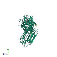
4mq0

X-ray diffraction
2.12Å resolution

Crystal structure of Parkia biglobosa seed lectin (PBL) in complex with methyl alpha D-mannopyranoside

Released:
Source organism: Parkia biglobosa
Entry authors: Teixeira CS, Rocha BAM, Silva HC, Bari AU, Barroso-Neto IL, Santiago MQ, Nagano CS, Nascimento KS, Debray H, Delatorre P, Cavada BS

Function and Biology Details

Biochemical function:
Biological process:
  • not assigned
Cellular component:
  • not assigned

Structure analysis Details

Assembly composition:
homo dimer (preferred)
PDBe Complex ID:
PDB-CPX-164827 (preferred)
Entry contents:
1 distinct polypeptide molecule
Macromolecule:
Parkia biglobosa lectin (PBL) Chain: A
Molecule details ›
Chain: A
Length: 446 amino acids
Theoretical weight: 47.61 KDa
Source organism: Parkia biglobosa
Structure domains: Jacalin-like lectin domain

Ligands and Environments

1 bound ligand:
No modified residues

Experiments and Validation Details

Entry percentile scores
X-ray source: LNLS BEAMLINE W01B-MX2
Spacegroup: C2221
Unit cell:
a: 77.139Å b: 86.659Å c: 127.857Å
α: 90° β: 90° γ: 90°
R-values:
R R work R free
0.26 0.258 0.293