


Function and Biology Details
Biochemical function:
Biological process:
Cellular component:
- not assigned
Sequence domain:
Structure analysis Details
Assembly composition:
monomeric (preferred)
Assembly name:
Endonuclease V (preferred)
PDBe Complex ID:
PDB-CPX-185437 (preferred)
Entry contents:
1 distinct polypeptide molecule
Macromolecule:
Endonuclease V
Molecule details ›
Chain: A
Length: 250 amino acids
Theoretical weight: 27.94 KDa
Source organism: Homo sapiens
Expression system: Escherichia coli
UniProt:
Sequence domains: Endonuclease V
Structure domains: archaeoglobus fulgidus dsm 4304 superfamily
Length: 250 amino acids
Theoretical weight: 27.94 KDa
Source organism: Homo sapiens
Expression system: Escherichia coli
UniProt:
- Canonical:
 Q8N8Q3 (Residues: 13-250; Coverage: 84%)
Sequence domains: Endonuclease V
Structure domains: archaeoglobus fulgidus dsm 4304 superfamily
Ligands and Environments
No bound ligands
No modified residues
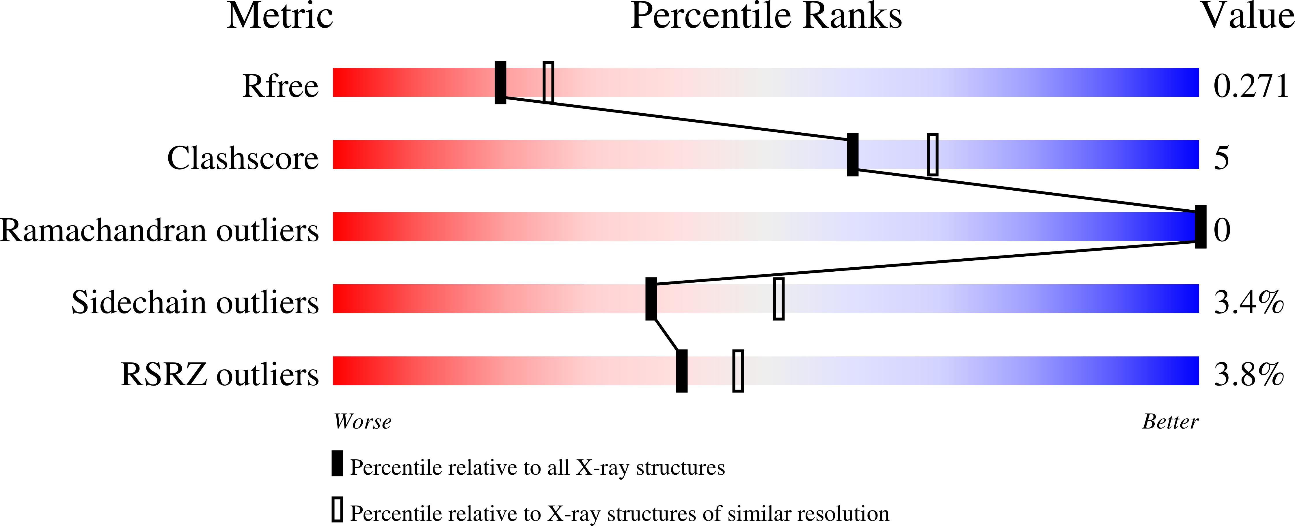
Experiments and Validation Details

X-ray source:
Nova high-flux-micro-focus sealed tube
Spacegroup:
P212121
Expression system: Escherichia coli