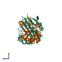
4u5y

X-ray diffraction
1.93Å resolution

Crystal Structure of the complex between the GNAT domain of S. lividans PAT and the acetyl-CoA synthetase C-terminal domain of S. enterica

Released:

Function and Biology Details

Structure analysis Details

Assembly composition:
hetero dimer (preferred)
PDBe Complex ID:
PDB-CPX-165208 (preferred)
Entry contents:
2 distinct polypeptide molecules
Macromolecules (2 distinct):
Acyl-CoA synthetase Chain: A
Molecule details ›
Chain: A
Length: 194 amino acids
Theoretical weight: 21.67 KDa
Source organism: Streptomyces lividans TK24
Expression system: Escherichia coli
Structure domains: Aminopeptidase
Acetyl-coenzyme A synthetase Chain: D
Molecule details ›
Chain: D
Length: 135 amino acids
Theoretical weight: 14.54 KDa
Source organism: Salmonella enterica subsp. enterica serovar Paratyphi B str. ATCC 10719
Expression system: Escherichia coli
Structure domains: GMP Synthetase; Chain A, domain 3

Ligands and Environments

1 bound ligand:
No modified residues

Experiments and Validation Details

Entry percentile scores
X-ray source: APS BEAMLINE 19-BM
Spacegroup: C2221
Unit cell:
a: 62.478Å b: 66.524Å c: 138.539Å
α: 90° β: 90° γ: 90°
R-values:
R R work R free
0.189 0.187 0.239
Expression system: Escherichia coli