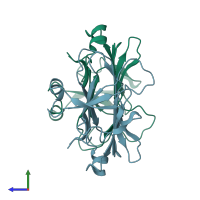
5b4n

X-ray diffraction
2.3Å resolution

Structure analysis of function associated loop mutant of substrate recognition domain of Fbs1 ubiquitin ligase

Released:

Function and Biology Details

Biochemical function:
  • not assigned
Biological process:
  • not assigned
Cellular component:
  • not assigned

Structure analysis Details

Assembly composition:
homo dimer (preferred)
PDBe Complex ID:
PDB-CPX-182375 (preferred)
Entry contents:
1 distinct polypeptide molecule
Macromolecule:
F-box only protein 2; F-box only protein 44 Chains: A, B
Molecule details ›
Chains: A, B
Length: 203 amino acids
Theoretical weight: 23.63 KDa
Source organisms: Expression system: Escherichia coli BL21(DE3)
UniProt:
  • Canonical: Q9H4M3 (Residues: 71-74, 99-102, 105-107, 110-133, 168-176, 194-208, 230-247; Coverage: 30%)
  • Canonical: Q80UW2 (Residues: 121-144, 149-150, 154-155, 181-214, 224-238, 254-274, 293-297; Coverage: 35%)
Gene names: FBG3, FBX30, FBX44, FBX6A, FBXO44, FBXO6A, Fbs1, Fbx2, Fbxo2
Sequence domains: F-box associated region
Structure domains: Galactose-binding domain-like

Ligands and Environments

No bound ligands
No modified residues

Experiments and Validation Details

Entry percentile scores
X-ray source: SPRING-8 BEAMLINE BL44XU
Spacegroup: P21
Unit cell:
a: 44.541Å b: 96.203Å c: 44.681Å
α: 90° β: 101.9° γ: 90°
R-values:
R R work R free
0.189 0.185 0.266
Expression system: Escherichia coli BL21(DE3)