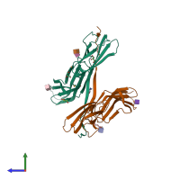
5c14

X-ray diffraction
2.8Å resolution

Crystal structure of PECAM-1 D1D2 domain

Released:
Source organism: Homo sapiens
Primary publication:
Structural basis for PECAM-1 homophilic binding.
Blood 127 1052-61 (2016)
PMID: 26702061

Function and Biology Details

Biochemical function:
  • not assigned
Biological process:
  • not assigned
Cellular component:
  • not assigned

Structure analysis Details

Assemblies composition:
monomeric
hetero tetramer (preferred)
PDBe Complex ID:
PDB-CPX-147672 (preferred)
Entry contents:
2 distinct polypeptide molecules
Macromolecules (2 distinct):
Platelet endothelial cell adhesion molecule Chain: A
Molecule details ›
Chain: A
Length: 212 amino acids
Theoretical weight: 24.53 KDa
Source organism: Homo sapiens
Expression system: Drosophila
UniProt:
  • Canonical: P16284 (Residues: 28-229; Coverage: 28%)
Gene name: PECAM1
Platelet endothelial cell adhesion molecule Chain: B
Molecule details ›
Chain: B
Length: 212 amino acids
Theoretical weight: 24.49 KDa
Source organism: Homo sapiens
Expression system: Drosophila
UniProt:
  • Canonical: P16284 (Residues: 28-229; Coverage: 28%)
Gene name: PECAM1

Ligands and Environments

1 modified residue:

Experiments and Validation Details

Entry percentile scores
X-ray source: APS BEAMLINE 21-ID-D
Spacegroup: I4122
Unit cell:
a: 104.01Å b: 104.01Å c: 281.834Å
α: 90° β: 90° γ: 90°
R-values:
R R work R free
0.239 0.238 0.279
Expression system: Drosophila