5fcg

X-ray diffraction
2.1Å resolution

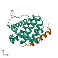
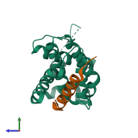
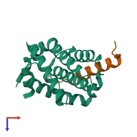
Crystal structure of Bcl-2 in complex with HBx-BH3 motif

Released:
Source organisms:
Primary publication:
Structural and biochemical analysis of Bcl-2 interaction with the hepatitis B virus protein HBx.
Proc Natl Acad Sci U S A 113 2074-9 (2016)
PMID: 26858413

Function and Biology Details

Structure analysis Details

Assembly composition:
hetero dimer (preferred)
PDBe Complex ID:
PDB-CPX-145309 (preferred)
Entry contents:
2 distinct polypeptide molecules
Macromolecules (2 distinct):
Apoptosis regulator Bcl-2 Chain: A
Molecule details ›
Chain: A
Length: 168 amino acids
Theoretical weight: 19.44 KDa
Source organism: Homo sapiens
Expression system: Escherichia coli BL21(DE3)
UniProt:
  • Canonical: P10415 (Residues: 1-207; Coverage: 70%)
Gene name: BCL2
Sequence domains:
Protein X Chain: C
Molecule details ›
Chain: C
Length: 26 amino acids
Theoretical weight: 3.16 KDa
Source organism: Hepatitis B virus
Expression system: Not provided
UniProt:
  • Canonical: Q99HR6 (Residues: 110-135; Coverage: 17%)
Gene name: X

Ligands and Environments

No bound ligands
No modified residues

Experiments and Validation Details

Entry percentile scores
X-ray source: SSRF BEAMLINE BL17U
Spacegroup: P21212
Unit cell:
a: 63.777Å b: 82.99Å c: 33.487Å
α: 90° β: 90° γ: 90°
R-values:
R R work R free
0.256 0.255 0.276
Expression systems:
  • Escherichia coli BL21(DE3)
  • Not provided