5lug

X-ray diffraction
1.7Å resolution

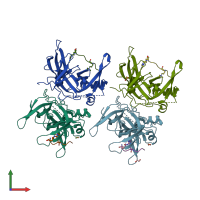
Crystal structure of human Spindlin-2B protein in complex with ART(M3L)QTA(2MR)KS peptide

Released:
Source organism: Homo sapiens
Entry authors: Srikannathasan V, Gileadi C, Talon R, Shrestha L, Kopec J, Szykowska A, Burgess-Brown N, Arrowsmith CH, Edwards AM, Bountra C, von Delft F, Oppermann U, Huber K

Function and Biology Details

Biochemical function:
  • not assigned
Biological process:
Cellular component:
  • not assigned

Structure analysis Details

Assembly composition:
hetero tetramer (preferred)
PDBe Complex ID:
PDB-CPX-159463 (preferred)
Entry contents:
2 distinct polypeptide molecules
Macromolecules (2 distinct):
Spindlin-2B Chains: A, B, C, D
Molecule details ›
Chains: A, B, C, D
Length: 222 amino acids
Theoretical weight: 25.51 KDa
Source organism: Homo sapiens
Expression system: Escherichia coli BL21(DE3)
UniProt:
  • Canonical: Q9BPZ2 (Residues: 44-258; Coverage: 83%)
Gene names: SPIN2, SPIN2B
Sequence domains: Spin/Ssty Family
Histone H3.1 Chains: E, F, G, H
Molecule details ›
Chains: E, F, G, H
Length: 10 amino acids
Theoretical weight: 1.22 KDa
Source organism: Homo sapiens
Expression system: Escherichia coli #1/H766
UniProt:
  • Canonical: P68431 (Residues: 2-11; Coverage: 7%)
Gene names: H3C1, H3C10, H3C11, H3C12, H3C2, H3C3, H3C4, H3C6, H3C7, H3C8, H3FA, H3FB, H3FC HIST1H3C, H3FD, H3FF, H3FH, H3FI, H3FJ, H3FK, H3FL, HIST1H3A, HIST1H3B, HIST1H3D, HIST1H3E, HIST1H3F, HIST1H3G, HIST1H3H, HIST1H3I, HIST1H3J

Ligands and Environments

2 bound ligands:
2 modified residues:

Experiments and Validation Details

Entry percentile scores
X-ray source: DIAMOND BEAMLINE I02
Spacegroup: P21
Unit cell:
a: 66.533Å b: 81.225Å c: 93.967Å
α: 90° β: 99.86° γ: 90°
R-values:
R R work R free
0.196 0.194 0.221
Expression systems:
  • Escherichia coli BL21(DE3)
  • Escherichia coli #1/H766