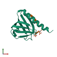
5u1m

X-ray diffraction
1.8Å resolution

Structure of the IRS-1 PTB Domain Bound to the Juxtamembrane Region of the Insulin Receptor

Released:

Function and Biology Details

Reaction catalysed:
ATP + a [protein]-L-tyrosine = ADP + a [protein]-L-tyrosine phosphate
Biochemical function:
Cellular component:
  • not assigned

Structure analysis Details

Assembly composition:
hetero dimer (preferred)
PDBe Complex ID:
PDB-CPX-138934 (preferred)
Entry contents:
2 distinct polypeptide molecules
Macromolecules (2 distinct):
Insulin receptor substrate 1 Chain: A
Molecule details ›
Chain: A
Length: 105 amino acids
Theoretical weight: 11.83 KDa
Source organism: Homo sapiens
Expression system: Escherichia coli K-12
UniProt:
  • Canonical: P35568 (Residues: 161-265; Coverage: 9%)
Gene name: IRS1
Sequence domains: PTB domain (IRS-1 type)
Structure domains: Pleckstrin-homology domain (PH domain)/Phosphotyrosine-binding domain (PTB)
Insulin receptor subunit beta Chain: B
Molecule details ›
Chain: B
Length: 10 amino acids
Theoretical weight: 1.09 KDa
Source organism: Homo sapiens
Expression system: Escherichia coli K-12
UniProt:
  • Canonical: P06213 (Residues: 991-999; Coverage: 1%)
Gene name: INSR

Ligands and Environments

No bound ligands
1 modified residue:

Experiments and Validation Details

Entry percentile scores
X-ray source: ELLIOTT GX-13
Spacegroup: P422
Unit cell:
a: 68.37Å b: 68.37Å c: 57.52Å
α: 90° β: 90° γ: 90°
Expression system: Escherichia coli K-12