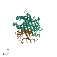
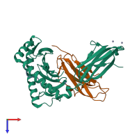
5vge

X-ray diffraction
2.6Å resolution

Crystal structure of HLA-C*07:02 in complex with RYR peptide

Released:

Function and Biology Details

Structure analysis Details

Assembly composition:
hetero trimer (preferred)
PDBe Complex ID:
PDB-CPX-145272 (preferred)
Entry contents:
3 distinct polypeptide molecules
Macromolecules (3 distinct):
HLA class I histocompatibility antigen, C alpha chain Chain: A
Molecule details ›
Chain: A
Length: 276 amino acids
Theoretical weight: 31.65 KDa
Source organism: Homo sapiens
Expression system: Escherichia coli
UniProt:
  • Canonical: P10321 (Residues: 26-300; Coverage: 80%)
Gene names: HLA-C, HLAC
Sequence domains:
Structure domains:
Beta-2-microglobulin Chain: B
Molecule details ›
Chain: B
Length: 100 amino acids
Theoretical weight: 11.88 KDa
Source organism: Homo sapiens
Expression system: Escherichia coli
UniProt:
  • Canonical: P61769 (Residues: 21-119; Coverage: 100%)
Gene names: B2M, CDABP0092, HDCMA22P
Sequence domains: Immunoglobulin C1-set domain
Structure domains: Immunoglobulins
Histone H3.1 Chain: C
Molecule details ›
Chain: C
Length: 9 amino acids
Theoretical weight: 1.03 KDa
Source organism: Homo sapiens
Expression system: Not provided
UniProt:
  • Canonical: P68431 (Residues: 41-49; Coverage: 7%)
Gene names: H3C1, H3C10, H3C11, H3C12, H3C2, H3C3, H3C4, H3C6, H3C7, H3C8, H3FA, H3FB, H3FC HIST1H3C, H3FD, H3FF, H3FH, H3FI, H3FJ, H3FK, H3FL, HIST1H3A, HIST1H3B, HIST1H3D, HIST1H3E, HIST1H3F, HIST1H3G, HIST1H3H, HIST1H3I, HIST1H3J

Ligands and Environments

1 bound ligand:
No modified residues

Experiments and Validation Details

Entry percentile scores
X-ray source: AUSTRALIAN SYNCHROTRON BEAMLINE MX1
Spacegroup: P65
Unit cell:
a: 99.67Å b: 99.67Å c: 77.914Å
α: 90° β: 90° γ: 120°
R-values:
R R work R free
0.166 0.163 0.221
Expression systems:
  • Escherichia coli
  • Not provided