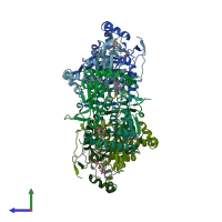
5yak

X-ray diffraction
2.3Å resolution

The crystal structure of human IYD Thr239 mutant with ligand 3-Fluorotyrosine (F-Tyr)

Released:
Source organism: Homo sapiens
Primary publication:
Redox control of iodotyrosine deiodinase.
Protein Sci 28 68-78 (2019)
PMID: 30052294

Function and Biology Details

Reaction catalysed:
(1a) L-tyrosine + NADP(+) + iodide = 3-iodo-L-tyrosine + NADPH
Biochemical function:
Biological process:
  • not assigned
Cellular component:
  • not assigned

Structure analysis Details

Assembly composition:
homo dimer (preferred)
PDBe Complex ID:
PDB-CPX-180158 (preferred)
Entry contents:
1 distinct polypeptide molecule
Macromolecule:
Iodotyrosine deiodinase 1 Chains: A, B, C, D, E, F
Molecule details ›
Chains: A, B, C, D, E, F
Length: 264 amino acids
Theoretical weight: 30.54 KDa
Source organism: Homo sapiens
Expression system: Escherichia coli
UniProt:
  • Canonical: Q6PHW0 (Residues: 32-289; Coverage: 89%)
Gene names: C6orf71, DEHAL1, IYD
Sequence domains: Nitroreductase family
Structure domains: NADH Oxidase

Ligands and Environments


Cofactor: Ligand FMN 6 x FMN
1 bound ligand:
No modified residues

Experiments and Validation Details

Entry percentile scores
X-ray source: NSLS BEAMLINE X25
Spacegroup: P61
Unit cell:
a: 105.079Å b: 105.079Å c: 300.202Å
α: 90° β: 90° γ: 120°
R-values:
R R work R free
0.185 0.183 0.226
Expression system: Escherichia coli