5i6s

X-ray diffraction
2.1Å resolution

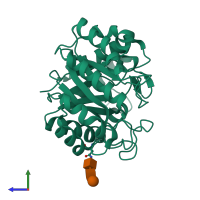
Crystal structure of an endoglucanase from Penicillium verruculosum

Released:
Source organism: Talaromyces verruculosus
Entry authors: Nemashklaov V, Vakhrusheva A, Tishchenko S, Gabdulkhakov A, Kravchenko O, Gusakov A, Sinitsyn A

Function and Biology Details

Reaction catalysed:
Endohydrolysis of (1->4)-beta-D-glucosidic linkages in cellulose, lichenin and cereal beta-D-glucans
Cellular component:
  • not assigned

Structure analysis Details

Assembly composition:
monomeric (preferred)
Assembly name:
PDBe Complex ID:
PDB-CPX-103762 (preferred)
Entry contents:
1 distinct polypeptide molecule
Macromolecules (2 distinct):
cellulase Chain: A
Molecule details ›
Chain: A
Length: 305 amino acids
Theoretical weight: 33.6 KDa
Source organism: Talaromyces verruculosus
UniProt:
  • Canonical: A0A1U7Q1U3 (Residues: 1-305; Coverage: 100%)
Sequence domains: Cellulase (glycosyl hydrolase family 5)
Structure domains: Glycosidases

Ligands and Environments

Carbohydrate polymer : NEW Components: NAG, BMA
No bound ligands
No modified residues

Experiments and Validation Details

Entry percentile scores
X-ray source: BRUKER AXS MICROSTAR
Spacegroup: P41212
Unit cell:
a: 83.819Å b: 83.819Å c: 89.044Å
α: 90° β: 90° γ: 90°
R-values:
R R work R free
0.22 0.218 0.26