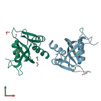
5ltu

X-ray diffraction
2.23Å resolution

Crystal Structure of NUDT4A- Diphosphoinositol polyphosphate phosphohydrolase 2

Released:
Source organism: Homo sapiens
Entry authors: Srikannathasan V, Nunez CA, Tallant C, Siejka P, Mathea S, Newman J, Strain-Damerell C, Elkins JM, Burgess-Brown N, Arrowsmith CH, Edwards AM, Bountra C, von Delft F, Huber K

Function and Biology Details

Reaction catalysed:
Diphospho-myo-inositol polyphosphate + H(2)O = myo-inositol polyphosphate + phosphate

Structure analysis Details

Assembly composition:
homo dimer (preferred)
PDBe Complex ID:
PDB-CPX-192758 (preferred)
Entry contents:
1 distinct polypeptide molecule
Macromolecule:
Diphosphoinositol polyphosphate phosphohydrolase 2 Chains: A, B
Molecule details ›
Chains: A, B
Length: 180 amino acids
Theoretical weight: 20.33 KDa
Source organism: Homo sapiens
Expression system: Escherichia coli
UniProt:
  • Canonical: Q9NZJ9 (Residues: 1-180; Coverage: 100%)
Gene names: DIPP2, HDCMB47P, KIAA0487, NUDT4
Sequence domains: NUDIX domain
Structure domains: Nucleoside Triphosphate Pyrophosphohydrolase

Ligands and Environments

1 bound ligand:
No modified residues

Experiments and Validation Details

Entry percentile scores
X-ray source: DIAMOND BEAMLINE I04-1
Spacegroup: C2
Unit cell:
a: 135.71Å b: 41.03Å c: 74.32Å
α: 90° β: 122.55° γ: 90°
R-values:
R R work R free
0.217 0.216 0.245
Expression system: Escherichia coli