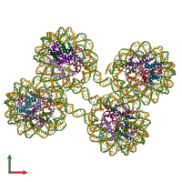
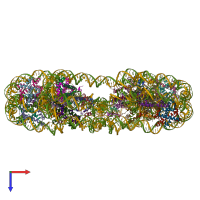
5oy7

X-ray diffraction
5.77Å resolution

Structure of the 4_601_157 tetranucleosome (P1 form)

Released:
Source organism: Xenopus laevis
Primary publication:
Capturing Structural Heterogeneity in Chromatin Fibers.
J Mol Biol 429 3031-3042 (2017)
PMID: 28893533

Function and Biology Details

Structure analysis Details

Assembly composition:
hetero 34-mer (preferred)
PDBe Complex ID:
PDB-CPX-119120 (preferred)
Entry contents:
4 distinct polypeptide molecules
2 distinct DNA molecules
Macromolecules (6 distinct):
Histone H2B 1.1 Chains: D, H, L, P, T, X, b, f
Molecule details ›
Chains: D, H, L, P, T, X, b, f
Length: 126 amino acids
Theoretical weight: 13.98 KDa
Source organism: Xenopus laevis
Expression system: Escherichia coli
UniProt:
  • Canonical: P02281 (Residues: 1-126; Coverage: 100%)
Sequence domains: Core histone H2A/H2B/H3/H4
Histone H3.2 Chains: A, E, I, M, Q, U, Y, c
Molecule details ›
Chains: A, E, I, M, Q, U, Y, c
Length: 135 amino acids
Theoretical weight: 15.3 KDa
Source organism: Xenopus laevis
Expression system: Escherichia coli
UniProt:
  • Canonical: P84233 (Residues: 2-136; Coverage: 99%)
Sequence domains: Core histone H2A/H2B/H3/H4
Histone H4 Chains: B, F, J, N, R, V, Z, d
Molecule details ›
Chains: B, F, J, N, R, V, Z, d
Length: 102 amino acids
Theoretical weight: 11.26 KDa
Source organism: Xenopus laevis
Expression system: Escherichia coli
UniProt:
  • Canonical: P62799 (Residues: 2-103; Coverage: 99%)
Sequence domains: Centromere kinetochore component CENP-T histone fold
Histone H2A type 1 Chains: C, G, K, O, S, W, a, e
Molecule details ›
Chains: C, G, K, O, S, W, a, e
Length: 130 amino acids
Theoretical weight: 14.11 KDa
Source organism: Xenopus laevis
Expression system: Escherichia coli
UniProt:
  • Canonical: P06897 (Residues: 1-130; Coverage: 100%)
Sequence domains:
DNA (619-MER) Chain: g
Molecule details ›
Chain: g
Length: 634 nucleotides
Theoretical weight: 194.77 KDa
DNA (619-MER) Chain: h
Molecule details ›
Chain: h
Length: 628 nucleotides
Theoretical weight: 195.07 KDa

Ligands and Environments

1 bound ligand:
No modified residues

Experiments and Validation Details

Entry percentile scores
X-ray source: SLS BEAMLINE X06SA
Spacegroup: P1
Unit cell:
a: 66.624Å b: 161.465Å c: 227.915Å
α: 78.94° β: 83.86° γ: 83.74°
R-values:
R R work R free
0.22 0.219 0.238
Expression system: Escherichia coli