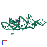
6fz0

X-ray diffraction
2.5Å resolution

Crystal structure of the metY SAM V riboswitch

Released:
Primary publication:
Structure and ligand binding of the SAM-V riboswitch.
Nucleic Acids Res 46 6869-6879 (2018)
PMID: 29931337

Function and Biology Details

Biochemical function:
  • not assigned
Biological process:
  • not assigned
Cellular component:
  • not assigned
Sequence domain:

Structure analysis Details

Assembly composition:
monomeric (preferred)
Assembly name:
PDBe Complex ID:
PDB-CPX-195913 (preferred)
Entry contents:
1 distinct RNA molecule
Macromolecule:
metY SAM V (53-MER) Chain: A
Molecule details ›
Chain: A
Length: 53 nucleotides
Theoretical weight: 17.13 KDa
Source organism: Candidatus Pelagibacter ubique HTCC1062
Expression system: Not provided
Sequence domains: SAM-V riboswitch

Ligands and Environments

1 modified residue:

Experiments and Validation Details

Entry percentile scores
X-ray source: DIAMOND BEAMLINE I03
Spacegroup: P4122
Unit cell:
a: 87.828Å b: 87.828Å c: 62.67Å
α: 90° β: 90° γ: 90°
R-values:
R R work R free
0.234 0.231 0.27
Expression system: Not provided