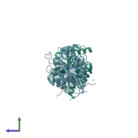
6hzl

X-ray diffraction
2.77Å resolution

Crystal structure of redox-inhibited phosphoribulokinase from Synechococcus sp. (strain PCC 6301), osmate derivative

Released:
Primary publication:
Crystal structure of phosphoribulokinase from Synechococcus sp. strain PCC 6301.
Acta Crystallogr F Struct Biol Commun 75 278-289 (2019)
PMID: 30950829

Function and Biology Details

Reaction catalysed:
ATP + D-ribulose 5-phosphate = ADP + D-ribulose 1,5-bisphosphate
Biochemical function:
Cellular component:
  • not assigned

Structure analysis Details

Assembly composition:
homo dimer (preferred)
Assembly name:
PDBe Complex ID:
PDB-CPX-102036 (preferred)
Entry contents:
1 distinct polypeptide molecule
Macromolecule:
Phosphoribulokinase Chains: A, B
Molecule details ›
Chains: A, B
Length: 333 amino acids
Theoretical weight: 37.76 KDa
Source organism: Synechococcus elongatus PCC 6301
Expression system: Escherichia coli BL21(DE3)
UniProt:
  • Canonical: A0A0H3K6J7 (Residues: 1-333; Coverage: 100%)
Gene names: prk, syc0567_d
Sequence domains: Phosphoribulokinase / Uridine kinase family

Ligands and Environments

1 bound ligand:
No modified residues

Experiments and Validation Details

Entry percentile scores
X-ray source: ESRF BEAMLINE ID29
Spacegroup: R32
Unit cell:
a: 141.432Å b: 141.432Å c: 206.563Å
α: 90° β: 90° γ: 120°
R-values:
R R work R free
0.236 0.235 0.259
Expression system: Escherichia coli BL21(DE3)